
Copper Nanoparticles Enhance Bactericidal Activity of 70% Ethanol Against Multidrug-Resistant Serratia marcescens.

Johan Insuasti-Cruz 1*, Juan Soto-Colina 1, Hugo Sánchez-Moreno 1, Segundo Hugo Calderón 1,
Naomi Rey-Moncayo 2
1Grupo de Investigación de Energías Alternativas y Ambiente, Facultad de Ciencias, Escuela Superior Politécnica de Chimborazo (ESPOCH), Panamericana Sur Km 1 ½, Chimborazo, EC060155; Ecuador
E-mail: juanfra3005@hotmail.com
E-mail: hugoj.sanchez@espoch.edu.ec;E-mail: hcalderon@espoch.edu.ec.
2Independent Researchers, Riobamba, 060104, Ecuador. E-mail: reynaomi45@gmail.com
*Correspondence: johan30ks@gmail.com, +593991459458
ABSTRACT
Copper nanoparticles (CuNPs) were synthesized by an adaptation of the chemical reduction method, using cupric sulfate pentahydrate (CuSO4·5H2O) and sodium borohydride (NaBH4) as a reducing agent, to combat bacterial resistance that leads to the proliferation of hospital infections caused by the bacterium Serratia marcescens through the potentiation of 70% ethyl alcohol (EA70) as a bactericidal agent through the addition of CuNPs. Characterization was performed with Fourier transform infrared spectroscopy (FTIR), UV-visible spectrophotometry, scanning electron microscopy (SEM), and energy dispersive spectroscopy (EDS), such that the CuNPs reached a diameter of 20-50 nm. The bactericidal activity was performed by inoculating the bacterium in Petri dishes with TM MEDIA Mueller Hinton agar, and its effectiveness was verified with susceptibility testing discs. The analysis was conducted at concentrations of 100, 300 and 500 mg/L; the current results have demonstrated that the lowest concentration shows a better inhibition halo, with a maximum of 13 mm, thus observing the synergism between both substances.
Keywords: Nanoparticles, antibiogram, inhibition, antimicrobial, nosocomial infection, synergism.

INTRODUCTION
Antiseptics are topical substances that eliminate microorganisms on the skin, mucous membranes, and wounds to prevent infectious diseases 1 . Bacterial resistance to these chemical agents is a problem that is scarcely considered worldwide, reducing the effectiveness of epidemic control and giving rise to microorganisms resistant to antibiotics and biocides in general 2 ,3 . This is generated by inadequate use or use of low concentrations of the same due to their ability to transfer antimicrobial resistance genes 4 . One of the most widely used antiseptics in the clinical and medical area is EA70, as it allows skin disinfection prior to blood draws and is used to perform prophylaxis of central venous catheters, highlighting that it kills up to 90% of bacteria when it remains on the skin for at least 2 minutes without drying after its application 5, 6 ,7 . EA70 fulfills a bactericidal function against certain vegetative microorganisms, whose mechanism of action is based on high-impact damage to the phospholipid layer of the cell membrane and to intracellular proteins, causing their denaturation 8 ,9 . Having portrayed this, it is essential to mention Serratia marcescens. This gram-negative bacillus belongs to the Enterobacteriaceae family as a bacteria of interest in the hospital environment since it has been shown that one of its reservoirs is the antiseptics used in this area, against which it has acquired resistance 10, 11 ,12 . This results in a significant health problem since the reduction in the bactericidal activity of these substances against this etiological agent allows it to be the cause of nosocomial infections, which are those contracted during a health care process in a health facility and whose main characteristic is that it was not present or in the incubation period before the patient's admission 13, 14 . Within these cases, this opportunistic microorganism has a prevalence of 1-2% and can cause urinary tract, central nervous system, eye and bloodstream infections, focusing its principal place of transmissibility in ICUs (Intensive Care Units) and newborn ICUs 15 ,16 ,17 . In the hospital context, a generally effective bactericidal agent is copper (Cu), which is used in the disinfection of high-contact surfaces; it is worth mentioning in this section that the United States Environmental Protection Agency has stated that Cu was the first element to act as an antimicrobial, also verifying its efficacy in the sterilization of wounds 18, 19 . Derivatives of this metal are not far behind, and among these are copper nanoparticles (CuNPs), which are tiny particles with effective bactericidal activity thanks to their surface-to-volume ratio, measuring 1-100 nanometers and covered by an interfacial layer composed of organic and inorganic molecules and ions 20 ,21 . The mechanism of action of CuNPs in bacterial cells is based on cell damage through the generation of reactive oxygen species (ROS), the release of Cu(II) ions, and the replacement or binding of native cofactors in metalloproteins. Also, Cu participates in innate immunity and can give way to the synthesis of ROS in the explosion reaction that occurs internally in phagocytes, increasing bactericidal activity in the process of bacterial phagocytosis 22, 23 . Despite this, there are strains of S. marcescens that are resistant to Cu (II), as these often originate from environments rich in heavy metals such as soil and water from mining deposits, for which they must adapt through different tolerance and elimination strategies such as intra and extracellular ion sequestration, the transformation of toxic metal species into non-toxic forms such as sulfides or metal oxides, development of permeability barriers, enzymatic detoxification, biosorption, precipitation, etc 24 ,25 . Given the acquired resistance of this commensal to the chemical agents described above, a solution that was considered viable to combat it is the addition of CuNPs to a specific antiseptic also used in hand disinfection, which in this case is EA70, taking into account that this last aspect reduces the rate of nosocomial infections by 40%, all in order to observe the synergistic behavior of both substances and thus increase their bactericidal power against this Enterobacteriaceae, this being the main focus of the present study, thus laying significant foundations in the fight against bacterial resistance to bactericides
MATERIALS AND METHODS
Reagents
CuSO4·5H2O 98% from the brand Sigma Aldrich, NaBH4 95% manufactured by Thermo Fisher Scientific, distilled water, gaseous nitrogen, EA70, sample of S. marcescens provided by the Laboratory of Biochemical and Microbiological Analysis of the Polytechnic School of Chimborazo, sterile saline solution 0.9%, BD Difco Nutrient Broth and TM MEDIA Mueller Hinton agar.
CuSO4·5H2O 98% from the brand Sigma Aldrich, NaBH4 95% manufactured by Thermo Fisher Scientific, distilled water, gaseous nitrogen, EA70, sample of S. marcescens provided by the Laboratory of Biochemical and Microbiological Analysis of the Polytechnic School of Chimborazo, sterile saline solution 0.9%, BD Difco Nutrient Broth and TM MEDIA Mueller Hinton agar.
Synthesis of Copper Nanoparticles
A modification of the chemical reduction method 27 synthesized the CuNPs. 0.5000 g of CuSO4·5H2O were weighed and filled to the mark in a 100 mL Florence flask with water treated with gaseous nitrogen. The solution was then sonicated with the Florence flask covered in the ultrasound equipment for 5 minutes. Subsequently, the contents were placed in a 250 mL beaker and agitated in the shaker for 30 min, uniformly adding 10 ml of a 0.8 M NaBH4 reducing mixture. The mixture sat for 48 hours, covered with aluminum foil. After this time, the samples were centrifuged for 5 min at 3000 rpm in a Digtor 21 C Ortoalresa centrifuge and washed with distilled water, 70% antiseptic alcohol and distilled water two more times using the same process. Lastly, the CuNPs were left to dry in an air circulation oven for 24 hours at 33 °C.
UV-Visible Spectrophotometry
The characterization of optical properties was carried out through scanning in a Thermo Fisher Scientific Evolution 220 spectrophotometer with an analysis range of 500 to 190 nm for the CuNPs and from 300 to 190 nm for the EA70 with CuNPs, where everything was operated with a resolution of 1 nm. The purpose of this equipment is to determine the wavelength at which the substances analyzed in this study absorb light significant way28 .
Fourier Transform Infrared Spectroscopy (FTIR)
The structural characterization of the copper nanoparticles was carried out by infrared spectroscopy with Fourier transform (FTIR), for which a JASCO FT/IR-4100 infrared spectrometer was used. This instrumental technique makes it possible to detect the rotation and vibration of molecules that respond to infrared radiation under a defined wavelength using characteristic peaks of the bonds of each compound in an analysis range of 4000 to 520 29
Scanning Electron Microscopy (SEM) with Energy Dispersive Spectroscopy (EDS)
The nanostructural characterization was carried out using a scanning electron microscope SEM, JSM-IT100LA, manufactured by JEOL, operated at 20.0 kV. This tool directs a beam of low-energy electrons toward the analyzed materials and observes its surface 30 . Coupled with this, the elemental composition analysis of the concentration and weight of CuNPs was carried out employing EDS 31 .
Evaluation of bactericidal activity
To evaluate bactericidal activity, a strain of S. marcescens was inoculated in BD Difco Nutrient Broth for enhancement and incubated at 37°C for 24 hours. Once this time had passed, an inoculum was prepared with sterile saline solution with 0.9% turbidity similar to scale 0.5 McFarland 108 CFU/ml and a massive inoculation was carried out with a sterile swab in quadruplicate in petri dishes with TM MEDIA Mueller Hinton agar 32 . Thermo Fisher susceptibility test discs with a diameter of 6 mm with 5, 7.5 and 10 mg CuNPs were placed in the culture medium, impregnated with a jaw press. Next, with the help of a micropipette, using the disc diffusion technique, 20 uL of 70%, 52.5% and 35% ethyl alcohol solutions were placed, respectively, and 20 uL of a prepared mixture of 100, 300 and 500 mg/L of CuNPs with EA70 33 . For the interpretation of results, the inhibition halo of each of the disks was measured with a conventional ruler.
RESULTS AND DISCUSSION
UV-Visible Spectrophotometry
The UV-visible spectrophotometry of the CuNPs gave a spectrum shown in Figure 1a, which peaked at 265 nm due to the absorption phenomenon of the surface plasmon, which originates from the joint oscillation of free electrons in the conduction band and which was stimulated by the incoming UV radiation; it is also important to note that the wavelength obtained is within a characteristic range, which is 200-300 nm, thus determining the presence of CuNPs 34 . Moreover, in the characterization of CuNPs with EA70, an absorption spectrum with an average peak of ≈193 nm was obtained for the three samples analyzed, which is observed in Figure 1b, and worth mentioning that the fact of adding alcohol as a solvent caused the absorption wavelength to decrease by approximately ≈72 nm, which is also known as hypochromic change, which occurs due to the π-π* stacking interaction when both substances react with each other 35 .

Figure 1. a) UV-visible spectrum of CuNPs; b) UV-visible spectrum of CuNPs + EA70 at different concentrations.
The FTIR spectrum shows a band characteristic of the stretching vibrations of the Cu-O bond, where the wavenumber associated with this typical peak is 598 cm-1 36 . Two peaks that are found at approximately 791 and 876 cm-1 indicate metal-oxygen (M-O) stretch vibrations; on the other hand, the bending and stretching vibrations that occur due to the moisture content on the surface of the CuNPs were determined by the peaks located approximately at 1655 and 3402 cm-1 37 . Bands between 800 and 1250 cm-1 could be part of the vibration modes of the remaining CuSO4·5H2O that did not react in the process 38 . Figure 2 shows the spectrum with the characteristics described in this section.

Figure 2. Fourier Transform Infrared Analysis Spectrum of CuNPs.
Scanning Electron Microscopy (SEM)
Figure 3 shows the nanoparticles microscopically at a scale of 1 um, with a diameter range of 20-50 nm and different polyhedral shapes, as well as considerable agglomeration 22 .

Figure 3. Analysis of CuNPs morphology using scanning electron microscopy.
Energy Dispersive Spectroscopy (EDS)
Samples were previously coated with gold through spray coating prior to observations to avoid the loading effect. The energy-dispersive spectroscopy presented a spectrum shown in Figure 4, where peaks of Cu are shown, confirming the presence of this metal. The mass ratio between Cu and O was 5.65% and 1.08%, respectively, while their atomic composition was 50.23% and 38%. Moreover, the presence of sulfur in 7.74% is a byproduct since the synthesis was made with CuSO4·5H2O. The presence of other metals, such as aluminum, are traces that arose during synthesis and do not intervene in the process.35

Figure 4. (a) Sample analysis area for (EDS); b) Spectrum of peaks of elements that make up the CuNPs sample
Bactericidal activity
When analyzing the results of the bactericidal activity of the different substances against S. marcescens, it was possible to observe that, in the case of CuNPs, no inhibition halos were obtained for 5 and 10 mg; however, for 7.5 mg, a confirmatory antimicrobial susceptibility test was performed again because the presence of a slight halo could be evidenced, where in this repetition of the procedure an inhibition zone of 8 mm was obtained. Additionally, in evaluating the bactericidal activity of ethyl alcohol (EA), inhibition halos of 6, 9 and 10 mm were obtained for 35%, 52.5% and 70%, respectively. Lastly, the bactericidal activity of EA70 in conjunction with CuNPs resulted in inhibition zones of 13, 10 and 9 mm for 100, 300 and 500 mg/L. Figure 5 and Figure 6 show the results photographically and statistically, respectively.
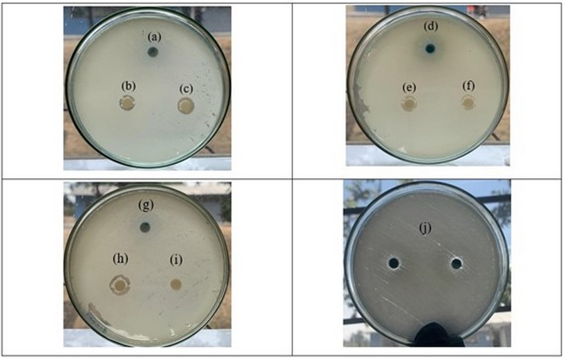
Figure 5. Comparison of the bactericidal activity of CuNPs, EA70 + CuNPs and EA at different concentrations against S. marcescens (a) 10mg of CuNPs, (b) 500 mg/L of EA70 + CuNPs c) EA70, (d) 7.5mg of CuNPs, (e) 300 mg/L of EA70 + CuNPs, (f) EA at 52.5% (g) 5mg of CuNP, (h), 100 mg/L of EA70 + CuNPs (i) EA at 35%, (j) 7.5 mg of CuNPs.

Figure 6. Zone of inhibition concerning the concentration of the bactericidal agents used. (a) CuNPs. (b) EA. (c) EA70 + CuNPs
S. marcescens was chosen as the study subject due to its well-known resistance to several biocides12 . The present research demonstrated that CuNPs alone exhibit significantly reduced bactericidal activity, possibly due to their stability being affected by the culture medium used for performing the antibiogram. It is essential to mention that stabilizing agents such as surface coating are necessary to avoid this. On the other hand, it was observed that the ideal concentration to potentiate EA70 by adding CuNPs was 100 mg/L or less; however, further studies are needed to confirm this. Therefore, it is possible to determine that increasing the concentration does not necessarily enhance the bactericidal effect. This occurs because high amounts of nanoparticles in the solvent tend to agglomerate with each other. It was considered that fewer agglomerations result in a larger available surface area, which induces more excellent ROS production and, thus an increase in their interaction with the bacterial membrane and the release of Cu (II) ions in the surrounding matrix. This phenomenon does not occur to a great extent in this case 39 .
The uniqueness of this study lies in its focus on the effectiveness of CuNPs combined with EA70 against S. marcescens, a bacterium that can be found in hospital antiseptics. The research results highlight the significant resistance of this bacterium to the mentioned substances, thus challenging conventional asepsis strategies. However, it is emphasized that adding CuNPs to EA70 shows promising potential to overcome this resistance and enhance the antiseptic's efficacy. These findings pave the way for new avenues of research in developing innovative solutions in the fight against resistant bacteria in clinical settings.
CONCLUSIONS
The characterization of the CuNPs by UV-visible spectrophotometry showed a characteristic peak at 265 nm due to the absorption phenomenon of the surface plasmon. Moreover, in the characterization of CuNPs with EA70, an absorption spectrum with an average peak of ≈193 nm was obtained for the three samples analyzed.
Fourier transform infrared spectroscopy provided a characteristic peak at 598 cm-1 , indicating a stretching vibration due to the presence of the Cu-O bond.
Scanning electron microscopy revealed the size of the CuNPs, whose length was approximately 20-50 nm, thus evidencing their nanometric size. The EDS was also performed, where peaks of Cu and O were observed.
CuNPs alone cannot significantly inhibit S. marcescens, as evidenced by the antimicrobial susceptibility test, given the formation of a small-diameter halo.
The EA showed no bactericidal power against S. marcescens at a concentration of 35% and slight effectiveness at a concentration of 52.5%.
An increased concentration of CuNPs in EA70 does not necessarily imply an improvement in bactericidal activity, as the agglomeration effect causes this. Using stabilizing agents in CuNPs is essential to ensure complete uniform dispersion and enhanced stability in the solution, which helps maintain their bactericidal activity and prevents particle aggregation.
Author Contributions: Johan Insuasti-Cruz: Writing – review & editing, Methodology, Formal analysis, Investigation, Conceptualization, Corresponding Author. Juan Soto-Colina: Writing – original draft, Methodology, Formal analysis, Investigation, Conceptualization. Hugo Sanchez-Moreno: Writing – original draft, Investigation, Conceptualization, Supervision. Segundo Hugo Calderón: Conceptualization, Supervision, Investigation. Naomi Rey-Moncayo: Editing, Validation, Supervision.
Acknowledgments: We sincerely thank Yolanda Buenaño, Geovanna Lobato, Mauricio Álvarez, Jefferson Bautista, and Pamela Morales, technicians in charge of the different laboratories of the Polytechnic School of Chimborazo, for their exceptional contributions throughout this research.
Funding: This research received no external funding.
Informed Consent Statement: Not applicable.
Conflicts of Interest: The authors declare no conflict of interest.
REFERENCES
1 González LL, Isabel Gutiérrez Pérez M, Eulalia Lucio-Villegas Menéndez M, Lluch NA, Luisa Morató Agustí M, Cachafeiro SP. Introducción a los antisépticos. Aten Primaria 2014; 46: 1–9.
2 van Dijk HFG, Verbrugh HA, Abee T, Andriessen JW, van Dijk HFG, ter Kuile BH et al. Resisting disinfectants. Communications Medicine 2022; 2: 6.
3 Zhelev G. Bacterial resistance to antiseptics and disinfectants – minireview. Bulg J Vet Med 2021; 24: 307–316.
4 Maillard J-Y, Pascoe M. Disinfectants and antiseptics: mechanisms of action and resistance. Nat Rev Microbiol 2023. doi:10.1038/s41579-023-00958-3.
5 Sarmah D. Alcohol Used as Disinfectant before Venipuncture does not Lead to Sample Haemolysis or Sample Dilution. JOURNAL OF CLINICAL AND DIAGNOSTIC RESEARCH 2016. doi:10.7860/JCDR/2016/15967.7245.
6 Zhang J, Wang B, Wang J, Yang Q. Ethanol locks for the prevention of catheter-related infection in patients with central venous catheter: A systematic review and meta-analysis of randomized controlled trials. PLoS One 2019; 14: e0222408.
7 del Río-Carbajo L, Vidal-Cortés P. Types of antiseptics, presentations and rules of use. Med Intensiva 2019; 43: 7–12.
8 León Molina J, Abad-Corpa E. Disinfectants and antiseptics facing coronavirus: synthesis of evidence and recommendations. Enferm Clin 2021; 31: S84–S88.
9 Lim K, Li WY, Dinata A, Ho ET. Comparing the antibacterial efficacy and functionality of different commercial alcohol-based sanitizers. PLoS One 2023; 18: e0282005.
10 Baráti-Deák B, Da Costa Arruda GC, Perjéssy J, Klupács A, Zalán Z, Mohácsi-Farkas C et al. Inhibition of Foodborne Pathogenic Bacteria by Excreted Metabolites of Serratia marcescens Strains Isolated from a Dairy-Producing Environment. Microorganisms 2023; 11: 403.
11 DOSSI C. MT, ESCALONA U. M, SERRANO A. C, SILVA D. MA, JULIET L. C, FERNÁNDEZ V. A et al. Serratia marcescens: Descripción de un brote de infección intrahospitalaria. Revista chilena de infectología 2002; 19: 262–266.
12 Lompo P, Heroes A-S, Agbobli E, Kühne V, Tinto H, Affolabi D et al. Bacterial Contamination of Antiseptics, Disinfectants and Hand Hygiene Products in Healthcare Facilities in High-Income Countries: A Scoping Review. Hygiene 2023; 3: 136–175.
13 Prabhu D, Rajamanikandan S, Amala M, Saritha P, Jeyakanthan J, Ramasamy P. Functional Characterization, Mechanism, and Mode of Action of Putative Streptomycin Adenylyltransferase from Serratia marcescens. Antibiotics 2022; 11: 1722.
14 Pujol M, Limón E. Epidemiología general de las infecciones nosocomiales. Sistemas y programas de vigilancia. Enferm Infecc Microbiol Clin 2013; 31: 108–113.
15 Khanna A. Serratia Marcescens - A Rare Opportunis - tic Nosocomial Pathogen and Measures to Limit its Spread in Hospitalized Patients. JOURNAL OF CLINICAL AND DIAGNOSTIC RESEARCH 2013. doi:10.7860/JCDR/2013/5010.2737.
16 Zivkovic Zaric R, Zaric M, Sekulic M, Zornic N, Nesic J, Rosic V et al. Antimicrobial Treatment of Serratia marcescens Invasive Infections: Systematic Review. Antibiotics 2023; 12: 367.
17 Cristina M, Sartini M, Spagnolo A. Serratia marcescens Infections in Neonatal Intensive Care Units (NICUs). Int J Environ Res Public Health 2019; 16: 610.
18 Montero DA, Arellano C, Pardo M, Vera R, Gálvez R, Cifuentes M et al. Antimicrobial properties of a novel copper-based composite coating with potential for use in healthcare facilities. Antimicrob Resist Infect Control 2019; 8: 3.
19 Crisan MC, Teodora M, Lucian M. Copper Nanoparticles: Synthesis and Characterization, Physiology, Toxicity and Antimicrobial Applications. Applied Sciences 2021; 12: 141.
20 Bhagat M, Anand R, Sharma P, Rajput P, Sharma N, Singh K. Review—Multifunctional Copper Nanoparticles: Synthesis and Applications. ECS Journal of Solid State Science and Technology 2021; 10: 063011.
21 Jayaraman A, Schweizer KS. Effective Interactions and Self-Assembly of Hybrid Polymer Grafted Nanoparticles in a Homopolymer Matrix. Macromolecules 2009; 42: 8423–8434.
22 Ma X, Zhou S, Xu X, Du Q. Copper-containing nanoparticles: Mechanism of antimicrobial effect and application in dentistry-a narrative review. Front Surg 2022; 9. doi:10.3389/fsurg.2022.905892.
23 Talebian S, Shahnavaz B, Nejabat M, Abolhassani Y, Rassouli FB. Bacterial-mediated synthesis and characterization of copper oxide nanoparticles with antibacterial, antioxidant, and anticancer potentials. Front Bioeng Biotechnol 2023; 11. doi:10.3389/fbioe.2023.1140010.
24 Díaz A, Marrero J, Cabrera G, Coto O, Gómez JM. Biosorption of nickel, cobalt, zinc and copper ions by Serratia marcescens strain 16 in mono and multimetallic systems. Biodegradation 2022; 33: 33–43.
25 Costa FS, Macedo MWFS, Araújo ACM, Rodrigues CA, Kuramae EE, de Barros Alcanfor SK et al. Assessing nickel tolerance of bacteria isolated from serpentine soils. Brazilian Journal of Microbiology 2019; 50: 705–713.
26 Kampf G, Löffler H, Gastmeier P. Hand Hygiene for the Prevention of Nosocomial Infections. Dtsch Arztebl Int 2009. doi:10.3238/arztebl.2009.0649.
27 Dang TMD, Le TTT, Fribourg-Blanc E, Dang MC. Synthesis and optical properties of copper nanoparticles prepared by a chemical reduction method. Advances in Natural Sciences: Nanoscience and Nanotechnology 2011; 2. doi:10.1088/2043-6262/2/1/015009.
28 Amaliyah S, Pangesti DP, Masruri M, Sabarudin A, Sumitro SB. Green synthesis and characterization of copper nanoparticles using Piper retrofractum Vahl extract as bioreductor and capping agent. Heliyon 2020; 6: e04636.
29 Faghihzadeh F, Anaya NM, Schifman LA, Oyanedel-Craver V. Fourier transform infrared spectroscopy to assess molecular-level changes in microorganisms exposed to nanoparticles. Nanotechnology for Environmental Engineering 2016; 1: 1.
30 Omidi M, Fatehinya A, Farahani M, Akbari Z, Shahmoradi S, Yazdian F et al. Characterization of biomaterials. In: Biomaterials for Oral and Dental Tissue Engineering. Elsevier, 2017, pp 97–115.
31 Kumari S, Singh BN, Srivastava P. Effect of copper nanoparticles on physico-chemical properties of chitosan and gelatin-based scaffold developed for skin tissue engineering application. 3 Biotech 2019; 9: 102.
32 Zeouk I, Ouedrhiri W, Sifaoui I, Bazzocchi IL, Piñero JE, Jiménez IA et al. Bioguided Isolation of Active Compounds from Rhamnus alaternus against Methicillin-Resistant Staphylococcus aureus (MRSA) and Panton-Valentine Leucocidin Positive Strains (MSSA-PVL). Molecules 2021; 26: 4352.
33 Ruparelia JP, Chatterjee AK, Duttagupta SP, Mukherji S. Strain specificity in antimicrobial activity of silver and copper nanoparticles. Acta Biomater 2008; 4: 707–716.
34 Rangasamy M, Gopal SK, Indhumathi A, Loganathan S, Manikandan S, Naresh R. Green Synthesis and Characterization of Copper Oxide Nanoparticles Using Tecoma Stans. J Pharm Res Int 2023; 35: 9–16.
35 Ni Y, Du S, Kokot S. Molecular spectroscopy and chemometrics: an analytical study of synergistic effects of drugs—interaction between fluoroquinolones and DNA. Analyst 2009; 134: 1840.
36 Mageshwari K, Sathyamoorthy R. Flower-shaped CuO Nanostructures: Synthesis, Characterization and Antimicrobial Activity. J Mater Sci Technol 2013; 29: 909–914.
37 Angeline Mary AP, Thaminum Ansari A, Subramanian R. Sugarcane juice mediated synthesis of copper oxide nanoparticles, characterization and their antibacterial activity. J King Saud Univ Sci 2019; 31: 1103–1114.
38 Gómez León MM, Román Mendoza LE, Castro Basurto FV, Maúrtua Torres DJ, Condori C, Vivas D et al. Nanopartículas de CuO y su propiedad antimicrobiana en cepas intrahospitalarias. Revista Colombiana de Química 2017; 46: 28–36.
39 Ameh T, Zarzosa K, Dickinson J, Braswell WE, Sayes CM. Nanoparticle surface stabilizing agents influence antibacterial action. Front Microbiol 2023; 14. doi:10.3389/fmicb.2023.1119550.
Received: April 22, 2024/ Accepted: May 25, 2024 / Published: June 15, 2024
Citation: Insuasti-Cruz J , Soto-Colina J, Sánchez-Moreno H, Hugo Calderón S, Rey-Moncayo N. Copper Nanoparticles Enhance Bactericidal Activity of 70% Ethanol Against Multidrug-Resistant Serratia marcescens. Bionatura journal 2024; 1 (2) 18. http://dx.doi.org/10.70099/BJ/2024.01.02.18
Additional information Correspondence should be addressed to johan30ks@gmail.com
Peer review information. Bionatura thanks anonymous reviewer(s) for their contribution to the peer review of this work using https://reviewerlocator.webofscience.com/
All articles published by Bionatura Journal are made freely and permanently accessible online immediately upon publication, without subscription charges or registration barriers.
Publisher's Note: Bionatura Journal stays neutral concerning jurisdictional claims in published maps and institutional affiliations.
Copyright: © 2024 by the authors. They were submitted for possible open-access publication under the terms and conditions of the Creative Commons Attribution (CC BY) license (https://creativecommons.org/licenses/by/4.0/).