
In vitro evaluation of the inhibitory capacity of three Trichoderma isolates on Ralstonia solanacearum

Jimmy Pico Rosado1*, Christopher Suárez Palacios1, Jessenia Jiménez Cumbicus1,
Ernesto Paredes Puga1, Gladys Sabando2, Liliana Andrade Olalla1
1 Instituto Nacional, de Investigaciones Agropecuarias, INIAP, Joya de los Sachas – Ecuador jimmy.pico@iniap.gob.ec.
1 Instituto Nacional, de Investigaciones Agropecuarias, INIAP, Joya de los Sachas – Ecuador christopher.suarez@iniap.gob.ec.
1 Instituto Nacional, de Investigaciones Agropecuarias, INIAP, Joya de los Sachas – Ecuador jessenia.jimenez@iniap.gob.ec.
1 Instituto Nacional, de Investigaciones Agropecuarias, INIAP, Joya de los Sachas – Ecuador ernesto.paredes@iniap.gob.ec.
1 Instituto Nacional, de Investigaciones Agropecuarias, INIAP, Joya de los Sachas – Ecuador liliana.andrade@iniap.gob.ec.
2 Instituto Superior Tecnológico General Eloy Alfaro, Joya de los Sachas – Ecuador gsabando@institutos.gob.ec.
* Correspondence: jimmy.pico@iniap.gob.ec
Available from. http://dx.doi.org/10.21931/BJ/2024.01.01.6
ABSTRACT
Bacterial wilt in bananas, caused by Ralstonia solanacearum or Moko, limits crop production and threatens Ecuador. This study evaluated Trichoderma isolates in laboratory conditions as an innovative alternative to ensure sustainability in banana production. The four R. solanacearum. isolates were obtained from banana plants exhibiting disease symptoms and were characterized through morphological and biochemical tests. Four treatments were evaluated: three isolates of fungi from the genus Trichoderma (Trichoderma viride, T. harzianum, T. asperellum) and one consisting of a combination of the three isolates above. The inhibitory capacity of the Trichoderma isolates on R. solanacearum colonies was measured. A completely randomized design with three replicates was used, and general linear and mixed models were employed, with qq-plot graphs for normality and residual plots for variance homogeneity.
Furthermore, a Fisher's LSD test was conducted at a significance level of α = 0.05. In the biochemical tests, the bacterial isolates exhibited specific characteristics of R. solanacearum in two bacterial isolates. In the inhibition tests, treatment four and treatment one (consortium of the three Trichoderma isolates and Trichoderma viride) showed the highest inhibitory potential, with 76.07% and 61.19%, respectively. The consortium of Trichoderma isolates demonstrated the highest inhibitory potential against R. solanacearum, with day 10 being the time with the highest percentage of inhibition (72.61%).
Keywords: Bacterial wilt, Ralstonia solanacearum, Trichoderma, inhibition
INTRODUCTION
In Ecuador, several species of Musaceae are cultivated, with bananas being the crop with the largest planted area, covering 165,080 hectares, followed by plantains with 145,501 hectares, and baby bananas or "oritos" with a cultivated area of 6,839 hectares (1–3). Plantains are a prominent export crop and a fundamental staple for the country's food security and employment generation (4). The central banana-producing provinces in Ecuador are Manabí, Santo Domingo, Esmeraldas, Guayas, Los Ríos, Orellana, Morona Santiago, Napo, and Sucumbíos (1).
In the banana belt of Ecuador, where varieties such as Dominico and Barraganete are grown, banana producers, mostly small-scale farmers, face significant challenges caused by phytopathogenic fungi and pests such as the black weevil (Cosmopolites sordidus), nematodes of the genera Pratylenchus and Helicotylenchus, and Black Sigatoka (Mycosphaerella fijiensis). One of the most significant challenges is Ralstonia solanacearum, known as "Moko," a bacterium that colonizes and obstructs the plant's vascular system, leading to wilting and, eventually, plant death (5). Despite being an essential crop for food security and the country's economy, this disease severely threatens plants.
Biological control has been adopted as part of integrated pest management to promote sustainable agriculture practices. In this context, fungi of the genus Trichoderma have shown effectiveness in controlling phytopathogenic fungi in different crops. Trichoderma spp., microscopic facultative anaerobic fungi, are naturally found in the soil and other environments with decomposing organic matter and plant residues (6,7).
This research aims to evaluate the potential of biological control using three Trichoderma isolates (T. viride, T. harzianum, T. asperellum) to inhibit the growth of Ralstonia solanacearum under laboratory conditions. The focus is reducing dependence on agrochemicals and minimizing negative environmental impacts. Additionally, using Trichoderma spp. as a biological control agent is expected to improve the plantain crop's productivity and mitigate problems caused by R. solanacearum in this critical sector. Through this research, we seek to provide scientific evidence of the effectiveness of biological control with Trichoderma spp. as a viable and sustainable alternative for managing R. solanacearum in the plantain crop. The results will strengthen integrated pest management strategies and promote more environmentally friendly agricultural practices, benefiting banana producers and food security in the country.
MATERIALS AND METHODS
The study was conducted at the Plant Protection Laboratory, Central Experimental Station of the Amazonia of the National Institute of Agricultural and Livestock Research (INIAP). This institution is located in San Carlos, La Joya de los Sachas Canton, Orellana Province, at 280 meters above sea level.
Obtaining Trichoderma isolates
The isolates identified as Trichoderma Viride, Trichoderma Harzianum, and Trichoderma Asperellum were provided by the Tropical Pichilingue Experimental Station and preserved in Eppendorf tubes with sterile distilled water, following the methodology described by (8). The main objective of this methodology is to maintain the viability, authenticity, and purity of the microorganisms.
Activation of Trichoderma isolates
The following steps were carried out to activate the Trichoderma isolates preserved in sterile water: the microtubes containing the Trichoderma strains were shaken in an isolation chamber. Then, 0.5 ml of each isolate was taken using a micropipette with previously sterilized tips. These samples were poured into the center of a Petri dish containing PDA (Potato Dextrose Agar) culture medium supplemented with lactic acid. The Petri dishes were incubated at a temperature of 27°C for 10 days, allowing the Trichoderma isolates to develop and grow actively.
Obtaining the pathogenic bacterial isolate
The following steps were followed for bacterial isolation from plant tissues with disease symptoms: portions of affected tissue were taken and superficially disinfected with a 4% sodium hypochlorite and 70% (v/v) alcohol solution. Subsequently, they were washed with sterile distilled water. Approximately 5 mm² each, tissue fragments were cut and macerated in a mortar using sterile purified water. The resulting suspension was spread on Petri dishes containing nutrient agar using a previously sterilized Drigalski spatula. After 48 hours of bacterial growth, a loopful of bacterial growth was transferred to a selective culture medium called "selective media from South Africa" (SMSA) modified. The Petri dishes were incubated at 30°C for 48 hours. During this time, typical colonies of slightly fluid reddish-purple color with a pink center were selected using criteria described by (9-10), and other relevant studies.
Characterization of pathogenic bacteria
Biochemical and physiological methods in plant microbiology studies were widely used to characterize the isolated bacteria based on the techniques described by Goszczynska and colleagues (11). These methods included Gram staining to determine cell morphology, the use of 3% KOH to identify Gram-negative or Gram-positive bacteria, the oxidase test to verify the production of the oxidase enzyme, the catalase test to detect the presence of the catalase enzyme, the nitrate reduction test to evaluate the bacteria's ability to reduce nitrate, growth at 41°C to determine thermotolerance, and salt tolerance by growth in media with NaCl concentrations of 1%, 2%, and 3%. Additionally, the fluorescence test was performed to detect the production of fluorescence, an essential criterion for identifying Ralstonia strains. These biochemical and physiological tests are fundamental for determining and characterizing isolated bacteria, as they provide essential information about their metabolic characteristics and ability to survive in different environmental conditions.
Conservation of bacteria
The methodology for conserving bacterial isolates described by (8) was used to maintain the bacteria's viability, authenticity, and purity over time. Purified bacterial isolates were cultured on Petri dishes with a nutrient agar medium. Then, sterile test tubes with nutrient agar medium were prepared and inclined at a 45° angle to allow the medium to solidify. The bacteria were streaked onto the inclined tubes and left to rest for one day at room temperature. Finally, the test tubes were preserved through freezing, ensuring the preservation of the bacterial isolates for future use.
Experiment management
To perform the inhibition tests, the methodology described by Andrade (11), was used with some modifications. First, in a laminar flow cabinet, enriched potato-dextrose-agar and nutrient agar media were dispensed in 90 mm Petri dishes. For the dual confrontation, a 5 mm diameter disc of each Trichoderma isolate and the bacterial isolates were placed opposite each other on the surface of the culture medium. As for the consortium confrontation, using a sterile punch, a 5 mm disc was set at three points of the Trichoderma isolates, and one disc was set at the center of the bacterial isolate that tested positive for Ralstonia. The Petri dishes were incubated at room temperature, and the interaction between the antagonistic organism and the pathogen was recorded every 24 hours. This recording allowed the observation and evaluation of the effects of Trichoderma isolates on the bacterial isolates, both in the dual confrontation and the consortium.
Inhibition tests of bacterial isolates
Tests were conducted to measure the inhibitory effect of three Trichoderma isolates (T. viride, T. harzianum, and T. asperellum) on two bacterial isolates positive for Ralstonia solanacearum. Five treatments were established: T1 (T. viride), T2 (T. harzianum), T3 (T. asperellum), T4 corresponding to the consortium of the three Trichoderma isolates, and T5 as the control, consisting of the unrestricted growth of bacterial isolates. A completely randomized design with three replications was used, with a Petri dish as the experimental unit (Table 1). This allowed for a systematic and controlled evaluation of the effect of different Trichoderma treatments on the bacterial isolates, providing precise information about the inhibitory potential of the Trichoderma isolates against R. solanacearum.
Study parameters
In evaluating inhibitory potential, the percentage of colony growth inhibition (PCGI) was determined using a modified formula based on the work of Suárez and Cabrales (12). This formula allowed for the precise calculation of the inhibition degree of bacterial colony growth in the presence of different Trichoderma treatments. In this way, the inhibitory effect of Trichoderma isolates on R. solanacearum isolates could be quantified objectively and comparatively.
Where:
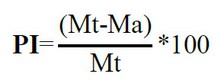
Ma: Ralstonia solanacearum, inhibited.
Mt: Mycelium of free growth of Ralstonia solanacearum (control).
Data analysis
In the statistical analysis, the Trichoderma isolates and the number of evaluations over time were considered fixed effects, while repetitions were treated as random. The data obtained were analyzed using the statistical software InfoStat version 2015. General linear and mixed models (13), were employed, and model assumptions were verified through qq-plot graphs for normality and residual plots against predicted values for variance homogeneity. Additionally, a Fisher's LSD test with a significance level of α= 0.05 was conducted to compare the means. To assess interactions between the antagonistic organism and the pathogen, the Image Tool program version 3.0, developed by Wilcox et al. (2002) in collaboration with the University of Texas, was used. Compatible with Windows 7, this program allowed for measurements using photographs as reference.
RESULTS
Characterization of Ralstonia solanacearum isolates
In the characterization of four bacterial isolates obtained from banana tissue exhibiting Moko symptoms, two bacteria (isolates 3 and 4) with distinctive morphological characteristics such as a reddish appearance, mucous texture, and positive biochemical tests for Ralstonia solanacearum were identified. These results are presented in Table 1, and the appearance of the bacteria can be observed in Figure 2.
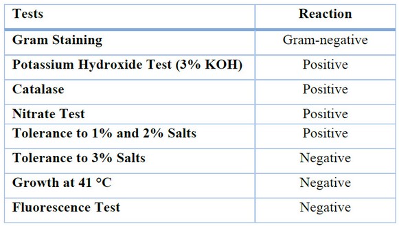
Table 1. Biochemical Test Results for Two Bacterial Isolates.
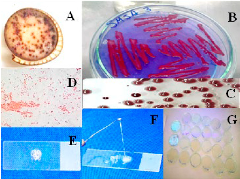
Figure 1. Response of the characterization of Ralstonia solanacearum. A; symptomatic tissue of the disease. B; Cultivation was performed in the SMSA culture medium. C; characteristics of the Ralstonia colony in SMSA medium. D; Gram stain (-). E; Catalase test (+). F; Potassium Hydroxide Test (+). G; Fluorescence test (-).
The results obtained in this research corroborate Torres's previous findings (14). who also found similar characteristics in bacterial colonies' morphology and polysaccharide production. Furthermore, the results of this research are consistent with Mendoza's reports (15). they were regarding the microscopic characteristics of Ralstonia solanacearum, such as its rod-shaped motile form and pink staining in the Gram stain using fuchsin. These results support the identification of the bacterium as Gram-negative, as determined by the 3% KOH test.
In summary, the results of the morphological and biochemical characterization of the bacterium in this research are consistent with previous findings and confirm the similarity of the isolated bacterium to Ralstonia. According to the study conducted by Pawaskar (16). it aligns with the results of identifying Ralstonia solanacearum based on morphological and biochemical characteristics, including Gram staining, KOH test, colony coloration, starch hydrolysis, and cellulose decomposition.
Inhibition tests
In Figure 2, it can be observed that the evaluated Trichoderma isolates exhibited a remarkable ability to inhibit the growth of the studied bacterial colonies. These results are consistent with those previously obtained by other researchers (17–19). It further supports the effectiveness of Trichoderma as a control agent. The treatments showed significant differences (P < 0.0001), indicating variability in the inhibitory potential of the different Trichoderma isolates. Specifically, treatment four, consisting of the consortium of the three Trichoderma isolates, and treatment one, showed the highest inhibitory potential with a percentage of colony growth inhibition of 76.07% and 61.19%, respectively. These values were statistically different from treatments two and three, which had inhibition percentages of 57.19% and 55.65%, respectively. The presented results demonstrate that the evaluated Trichoderma isolates can inhibit the growth of the studied bacterial colonies, with the consortium of the three isolates and an individual isolate being the most effective in this inhibition.
Means with a typical letter are not significantly different (p > 0.05)
Figure 2. Percentage of inhibition of Trichoderma isolates on Ralstonia sp. isolates (T1 Trichoderma viride, T2 Trichoderma harzianum, T3 Trichoderma asperellum, and T4 consortium of three Trichoderma isolates).
When analyzing the effect of days on inhibition, significant differences in days are observed (P < 0.0118), with day 10 achieving the highest percentage (72.61%), being statistically different from days six and two, which reached the lowest percentage (57.73% and 57.45%, respectively) (Figure 3).
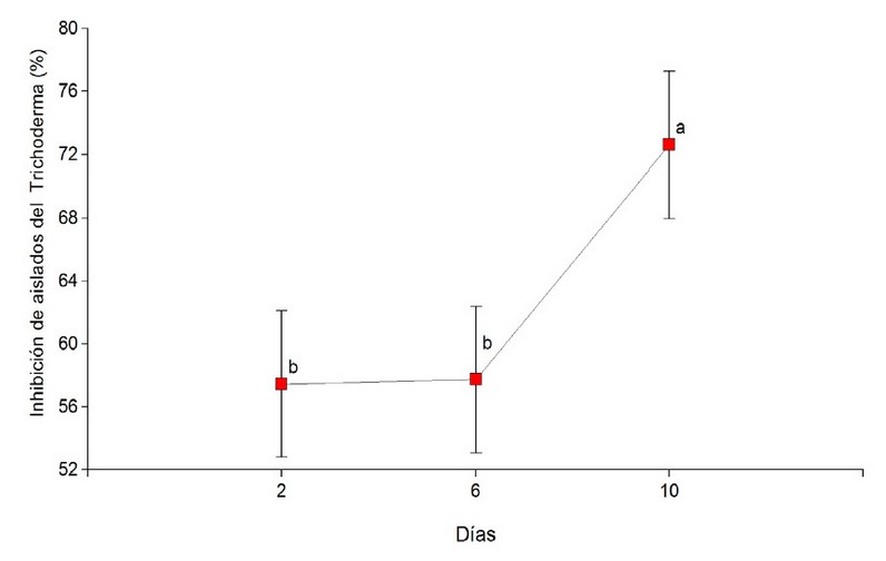
Means with a common letter are not significantly different (p > 0.05).
Figure 3. Percentage of inhibition of Trichoderma genus isolates on potential Ralstonia solanacearum isolates over time.
DISCUSSION
The in-depth analysis of the data presented, when compared to those of other authors, reveals that the results obtained in this research are consistent with previous studies conducted by Alvarez (20), Yendyo (21) and Goszczynska (22). These studies have demonstrated that Trichoderma genus isolates possess a significant antibiotic effect due to the production of thermostatic metabolites. These metabolites have been associated with an increase in root collar height and diameter, which correlates with the results obtained in this research regarding the inhibitory capacity of the consortium of Trichoderma isolates.
Furthermore, a study by other researchers (23-24) (25) found that applying metabolites from T. harzianum resulted in maximum inhibition of the soil bacterial population and reduced disease severity. These findings are similar to the results obtained in this research, where bacterial inhibition was observed starting from the tenth day after the application of Trichoderma isolates.
On the other hand, the findings of Ceballos (7), Align (26) and Sutarman (27), support the results of this research by demonstrating that crude extracts of Trichoderma sp. have highly significant effects on the in vitro inhibition of colonies of different species of R. solanacearum. These results strengthen the evidence of Trichoderma's antibacterial activity against the bacterium R. solanacearum.
According to the study by Khan (28), it aligns with the results obtained in this study, revealing that Trichoderma spp. Isolates significantly inhibited bacterial growth and cell damage by R. solanacearum.
CONCLUSIONS
This scientific study has significantly advanced our understanding of the interaction between Trichoderma isolates and Ralstonia solanacearum under laboratory conditions, as suggested in the article's title: "In vitro Evaluation of the Inhibitory Capacity of Three Trichoderma Isolates on Ralstonia solanacearum." Based on our results and concerning the objectives set forth, we have conclusively demonstrated that Trichoderma, especially the consortium of the three Trichoderma isolates and the T. viride isolate, possess a remarkable inhibitory potential against R. solanacearum. These findings strongly support using Trichoderma as an effective biological control agent in managing this phytopathogenic bacterium in banana crops. Furthermore, our results pave the way for future research and applications in this field, potentially significantly benefiting farmers and promoting sustainable and safe agricultural practices for food production.
REFERENCES
.1. ESPAC. Encuesta de Superficie y Producción Agropecuaria Continua [Internet]. Instituto Nacional de Estadistica y Censos; 2020 [citado 3 de febrero de 2021] p. T34. Disponible en: https://www.ecuadorencifras.gob.ec/estadisticas-agropecuarias-2/
2. INEC IN de E y C. Superficie, según producción y ventas de orito (fruta fresca) por región y provincia en Ecuador. Instituto Nacional de Estadística y Censo; 2020.
3. SIPA S de IPA del E. CIFRAS AGROPRODUCTIVAS [Internet]. Ministerio de Agricultura y Ganadería; 2020. Disponible en: http://sipa.agricultura.gob.ec/index.php/cifras-agroproductivas
4. Ordoñez J, Cevallos HV, Barrezueta-Unda S. Análisis de rentabilidad económica del plátano (Musa balbisiana AAB Simmond) en el sitio Río Negro, provincia El Oro. Revista Metropolitana de Ciencias Aplicadas. 2019;2(2):160-70.
5. Ramírez M, Neuman BW, Ramírez CA. Bacteriophages as promising agents for the biological control of moko disease (Ralstonia solanacearum) of banana. Biological Control. 2020;149:104238.
6. Suárez C., Remache N., Pico J., Paredes E., Jiménez J, Andrade L., et al. Aislamiento y evaluación de cepas nativas de Trichoderma spp., como promotor de desarrollo radicular. CU [Internet]. 27 de mayo de 2023 [citado 31 de mayo de 2023];16(42). Disponible en: https://ojs.unemi.edu.ec/index.php/cienciaunemi/article/view/1719
7. Ceballos G, Álvarez E, Bolaños MM. Reducción de poblaciones de Ralstonia solanacearum raza 2 (Smith) en plátano (Musa AAB Simmonds) con aplicación de extractos de Trichoderma sp.(Alexopoulus y Mims) y bacterias antagonistas. Acta agrónomica. 2014;63(1):80-7.
8. Maridueña-Zavala MG, Quevedo A, Aguaguiña K, Serrano L, Sosa del Castillo D. Colección de cultivos microbianos CIBE (CCM-CIBE): Una colección para la investigación. Bionatura. 2021;6:1664-8.
9. Denny T. II. Gram negative bacteria, F. Ralstonia. Laboratory guide for identification of plant pathogenic bacteria. 2001;151-74.
10. Schaad NW, Jones JB, Chun W. Laboratory guide for the identification of plant pathogenic bacteria. American Phytopathological society (APS press); 2001.
11. Andrade-Hoyos P, Luna-Cruz A, Osorio-Hernández E, Molina-Gayosso E, Landero-Valenzuela N, Barrales-Cureño HJ. Antagonismo de Trichoderma spp. vs hongos asociados a la marchitez de chile. Revista mexicana de ciencias agrícolas. 2019;10(6):1259-72.
12. Suárez LY, Cabrales CP. Identificación de especies de cepas nativas de Trichoderma sp. y Bacillus sp. y evaluación de su potencial antagonista in vitro frente al hongo fitopatógeno nativo Moniliophthora roreri en el departamento de Norte de Santander. Respuestas. 2008;13(1):45-56.
13. Di Rienzo J, Casanoves F, Gonzales L, Tablada E, Díaz M, Robledo C, et al. Estadistica para las Ciencias Agropecuarias. Séptima Edición ed. Cordoba Argentina, Editorial Brujas p. 2008;
14. Torres-González C, Casas M, Díaz Ortiz JE. Manejo de Ralstonia Solanacearum raza 2 a través de productos químicos y biológicos. Iteckne. 2013;10(2):217-23.
15. Mendoza Andrade OA. Caracterización filogenética molecular de secuencias sobre genes de siete cepas de Ralstonia solanacearum. 2021;
16. Pawaskar J, Joshi M, Navathe S, Agale R, Sawant Konkan Krishi Vidyapeeth B. Physiological and biochemical characters of ralstonia solanacearum. International Journal of Research in Agricultural Sciences. 2014;1(6):2348-3997.
17. Narasimha Murthy K, Nirmala Devi D, Srinivas C. Efficacy of Trichoderma asperellum against Ralstonia solanacearum under greenhouse conditions. Annals of Plant Sciences. 2013;2(9):342-50.
18. Konappa N, Krishnamurthy S, Siddaiah CN, Ramachandrappa NS, Chowdappa S. Evaluation of biological efficacy of Trichoderma asperellum against tomato bacterial wilt caused by Ralstonia solanacearum. Egyptian Journal of Biological Pest Control. 2018;28:1-11.
19. Mohamed BF, Sallam NM, Alamri SA, Abo-Elyousr KA, Mostafa YS, Hashem M. Approving the biocontrol method of potato wilt caused by Ralstonia solanacearum (Smith) using Enterobacter cloacae PS14 and Trichoderma asperellum T34. Egyptian Journal of Biological Pest Control. 2020;30:1-13.
20. CIAT GCI, Álvarez E, D CIAT P, Llano G. Actividad antagónica de bacterias biocontroladoras y filtrados de Trichoderma spp. contra Ralstonia solanacearum en plátano.
21. Yendyo S, Ramesh G, Pandey BR. Evaluation of Trichoderma spp., Pseudomonas fluorescens and Bacillus subtilis for biological control of Ralstonia wilt of tomato. F1000Research. 2017;6.
22. Goszczynska T, Serfontein J, Serfontein S. Introduction to practical phytobacteriology: a manual for phytobacteriology by SAFRINET, SDC Switzerland. 83 p. Disponible en línea: https://www researchgate net/publication/237021880_Introduction_to_Practical _Phytobacteriology_A_manual_for_phytobacteriology. 2000;
23. Guo Y, Fan Z, Yi X, Zhang Y, Khan RAA, Zhou Z. Sustainable Management of Soil-Borne Bacterium Ralstonia solanacearum In Vitro and In Vivo through Fungal Metabolites of Different Trichoderma spp. Sustainability. 2021;13(3):1491.
24. Yan L, Khan RAA. Biological control of bacterial wilt in tomato through the metabolites produced by the biocontrol fungus, Trichoderma harzianum. Egyptian Journal of Biological Pest Control. 2021;31(1):1-9.
25. Rahman M, Borah SM, Borah PK, Bora P, Sarmah BK, Lal MK, et al. Deciphering the antimicrobial activity of multifaceted rhizospheric biocontrol agents of solanaceous crops viz., Trichoderma harzianum MC2, and Trichoderma harzianum NBG. Frontiers in Plant Science. 2023;14.
26. Alelign S. Evaluation of the efficacy of Trichoderma and Pseudomonas species against bacterial wilt (Ralstonia isolates) of tomato (Lycopersicum spp.). 2020;
27. Sutarman S, Jalaluddin AK, Li’aini AS, Prihatiningrum AE. Characterizations of Trichoderma sp. and its effect on Ralstonia solanacearum of tobacco seedlings. Jurnal Hama dan Penyakit Tumbuhan Tropika. 2021;21(1):8-19.
28. Khan RAA, Najeeb S, Mao Z, Ling J, Yang Y, Li Y, et al. Bioactive secondary metabolites from Trichoderma spp. against phytopathogenic bacteria and root-knot nematode. Microorganisms. 2020;8(3):401.
Received: 9 October 2023/ Accepted: 15 January 2024 / Published:15 February 2024
Citation. Pico Rosado J, Suárez Palacios C, Jiménez Cumbicus J, Paredes Puga E, Sabando G, Andrade Olalla L. In vitro evaluation of the inhibitory capacity of three Trichoderma isolates on Ralstonia solanacearum. Revis Bionatura 2024; 1 (1) 6. http://dx.doi.org/10.21931/RB/2024.01.01.6
Additional information Correspondence should be addressed to jimmy.pico@iniap.gob.ec
Peer review information. Bionatura thanks anonymous reviewer(s) for their contribution to the peer review of this work using https://reviewerlocator.webofscience.com/
All articles published by Bionatura Journal are made freely and permanently accessible online immediately upon publication, without subscription charges or registration barriers.
Publisher's Note: Bionatura stays neutral concerning jurisdictional claims in published maps and institutional affiliations.
Copyright: © 2024 by the authors. They were submitted for possible open-access publication under the terms and conditions of the Creative Commons Attribution (CC BY) license (https://creativecommons.org/licenses/by/4.0/).