Electrospun fibers based on chitosan-carbon materials for electrochemical enzyme biosensor: advances and prospects to commercialization

Dulexy Solano-Orrala 1, Nayeli Gomez Castillo 1 , Christian Narváez-Muñoz 2,
Camilo Zamora-Ledezma 1,*
Camilo Zamora-Ledezma 1,*
¹ Bioengineering & Regenerative Medicine Research Group (Bio-ReM), Escuela de Ingeniería, Arquitectura y Diseño (EIAD), Universidad Alfonso X el Sabio (UAX), Avenida de la Universidad 1, 28691 Villanueva de la Cañada, Madrid, España.
² Departamento de Ciencias de la Energía y Mecánica, Universidad de las Fuerzas Armadas (ESPE), Sangolquí 171103, Ecuador.
² Departamento de Ciencias de la Energía y Mecánica, Universidad de las Fuerzas Armadas (ESPE), Sangolquí 171103, Ecuador.
* Corresponding author: Camilo Zamora-Ledezma, Email: camilza@uax.es
ABSTRACT
Electrospinning is a tunable technique for fabricating nanofibrous materials with exceptional properties for biosensing. The high surface area, interconnected porosity, and loading capacity of these nanofibers create an ideal microenvironment for enhancing sensor performance. This article focuses on composite platforms that synergistically combine the biocompatibility of chitosan with the improved electrical properties of carbon-based materials to develop highly sensitive and selective biosensors. Despite promising results, significant challenges hinder their commercial translation, including long-term enzyme stability, matrix interference from complex samples, fabrication protocols, and performance validation in real-world applications. Accordingly, this work critically assesses recent advancements in electrospun chitosan-carbon electrochemical enzyme biosensors, analyzes key technical hurdles, and discusses immobilization strategies crucial for achieving the reproducibility and scale required for industrial adoption.
Keywords: biosensor; electrospinning; chitosan; carbon; graphene; nanofiber.
INTRODUCTION
Rooted in their unique properties, the advent of nanomaterials has permanently reshaped the approach to biosensing. Nanofibrous scaffolds are particularly promising due to their high surface area to volume ratio. Among the techniques available for their fabrication, electrospinning has emerged as a predominant method. Compared to alternatives such as melt blowing or force spinning, electrospinning offers a superior combination of operational simplicity, cost-effectiveness, and unparalleled control over producing continuous nanofibers with tunable morphology and composition. This versatility allows for the integration of diverse functional materials beyond the base polymer, making it a powerful tool for creating sophisticated sensor platforms 1.
A highly effective strategy in this matter involves the development of nanocomposites that leverage the unique natural properties of distinct materials. In this context, chitosan, a natural biopolymer, is an ideal choice for the primary matrix. Its inherent biocompatibility, non-toxicity, and abundance of amine and hydroxyl functional groups provide a remarkable substrate for the stable immobilization of biological recognition elements. To enhance the functionality of this biocompatible scaffold, particularly for electrochemical applications, conductive materials such as graphene and its derivatives can be incorporated in the electrospun matrix. Renowned for their extraordinary mechanical strength and exceptional electrical conductivity, graphene-based materials act as a conductive backbone, facilitating efficient electron transfer and enhancing the signal transduction necessary for high-sensitivity detection2.
The integration of these components via electrospinning yields multifunctional biosensing platforms. Therefore, providing a unique synergy of high surface area, adjustable porosity, and enhanced conductivity collectively improves sensitivity and selectivity. However, despite these significant advantages, the translation of these platforms from laboratory to real-world application is hindered by several critical challenges. Major issues include achieving consistent conductivity throughout the mat, ensuring the long-term stability and activity of immobilized enzymes, overcoming batch-to-batch fabrication variability, and validating sensor performance in complex biological or environmental samples.
Therefore, this review article critically examines the state of the art in biosensors based on electrospun chitosan graphene composites. This work analyzes the synergistic interplay between these materials, evaluates current fabrication and biomolecule immobilization strategies, and discusses the primary scientific and engineering challenges that must be addressed to enable their practical implementation.
Electrospinning, an advanced fabrication technique
Electrospinning is an advanced fabrication technique widely recognized for producing continuous fibrous structures at the nanoscale. This bottom-up technique relies on electrostatic forces to elongate a polymeric solution, resulting in the formation of nanofibers3. The process involves applying a high voltage electrostatic field between two electrodes: a nozzle connected to a syringe and a metallic collector plate. This applied voltage overcomes the solution’s surface tension, causing the droplet at the nozzle tip to deform into a cone-shaped structure known as a Taylor cone4. Once a critical point is reached, a fine jet of the polymer solution erupts from the cone’s apex. As this jet travels toward the collector, the solvent evaporates, and the jet undergoes significant stretching and thinning. The solidified, continuous fiber is then deposited onto the collector, forming a non-woven, porous fibrous mat 5. Conventionally, electrospinning uses polymer solutions in organic solvents, with common polymers including nylon, polyester, and polylactic acid. Recent advancements have incorporated a broader range of materials, including ceramics, metals, and other inorganic and organic substances, as well as biological macromolecules like proteins and genes6. This ability to create composite materials opens the door to a new generation of advanced materials with industrial opportunities in biomedicine, filtration, energy, and environmental science.
Electrospun materials exhibit several highly desirable characteristics, including a high specific surface area, a highly interconnected porous structure, and high loading capacity and encapsulation efficiency7. The exceptionally high surface area to volume ratio of these scaffolds is particularly advantageous, as it enhances cell attachment, increases drug loading capacity, and improves mass transfer properties8. Therefore, they can serve as protective matrices for sensitive molecules like proteins and genes, leading to stable formulations with sustained release capabilities, which makes them highly valuable as drug carriers and as porous, biodegradable scaffolds that provide structural support for cells in tissue engineering 7. Consequently, they are widely used in medical prostheses and advanced wound healing solutions. Beyond biomedicine, applications extend to specialized textiles, such as waterproof breathable fabrics, and agriculture, where nanofiber webs carrying pesticides can be formed directly on plants to protect them from insects without the need for direct spraying.
The morphology and properties of the end material are highly dependent on the process parameters. Fundamental factors such as the viscosity and electrical conductivity of the polymer solution, as well as the applied voltage, directly influence the diameter of the fibers, which can range from micrometers to a few nanometers, but also control the pore size and spatial arrangement of the fibers, facilitating the formation of tailored nanofiber networks9–12. By carefully regulating these operational conditions, it is possible to fabricate membranes with diverse morphologies and properties for specific applications. This tunability enables the creation of specialized materials with unique properties, ideal for a wide range of applications.
Electrospun biosensing matrices
A particularly promising application of electrospun nanofibers is in the field of biosensing. As seen in Figure 1, a biosensor is an analytical device that combines a biological recognition element with a physicochemical transducer to detect a specific substance, or analyte 13,14. They are typically classified based on their transduction mechanism, such as electrochemical, optical, or thermometric systems15 The biomolecules responsible for recognition are generally immobilized on the surface of the detection component. When they interact with the target analyte, they generate physicochemical changes that the transducer measures as signals.
Biosensors are widely used in disease diagnostics, food safety, and environmental monitoring because conventional analytical methods, while powerful, are not always practical for rapid, accessible, or continuous monitoring. However, the practical application of typical biosensors is often limited by issues of low surface area, detection limits, and slow analyte diffusion. Electrospun nanofibers offer a great solution to these challenges. The nanoscale dimensions of the fibers create a massive surface area, making the transducer materials more active and accessible to the analyte. With diameters ranging from tens to hundreds of nanometers, electrospun fibers fall perfectly within this ideal regime, creating an entangled network that enhances sensor performance. To develop functional biosensors, nanofibers are often integrated with various active materials16. For instance, carbon-based materials like graphene oxide, carbon nanotubes, and carbon dots are widely used due to their high electrical conductivity and ease of functionalization 2. To illustrate, composites of conductive polymers, such as polyaniline (PANI), with carbon nanotubes can be electrospun to form highly sensitive electrochemical biosensors for glucose and other biomarkers. 17
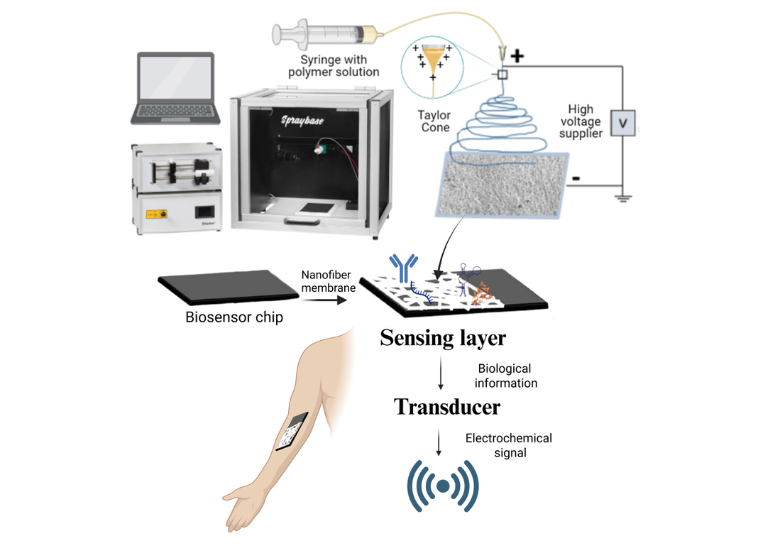
Figure 1. Schematic representation of a biosensor device based on electrospun fibers. The diagram shows the electrospinning process from a polymer solution syringe (Taylor cone formation and nanofiber membrane deposition), integration of the nanofiber membrane into a biosensor chip, and detection through a sensing layer that converts biological information into an electrochemical signal transmitted by the transducer.
Electrospun carbon-chitosan biosensors
Natural biopolymers like chitosan, silk fibroin, and hyaluronic acid are often blended with other polymers to enhance the biocompatibility and functionality of electrospun biosensors. Among these, chitosan stands out as a biocompatible, biodegradable, non-toxic, and hydrophilic material, making it safe for medical and environmental applications. Chitosan is a polysaccharide derived from the deacetylation of chitin, which is the second most abundant polysaccharide in nature and a primary component of crustacean exoskeletons. Its functional groups (amino and hydroxyl) enable chemical modification and direct conjugation with sensing elements like enzymes or antibodies, enhancing specificity. These properties, combined with their cost-effectiveness and rapid degradability, allow the creation of sensitive, selective, and stable biosensors for a wide range of analytes, including glucose, hydrogen peroxide, uric acid, and hormones such as acetylcholine18–22.
However, chitosan presents several challenges when used in its pure form, particularly during the electrospinning process. Due to its poor chain entanglement, it’s inherently difficult to electrospin alone, often leading to a non-uniform fiber morphology. Consequently, it is frequently blended with synthetic polymers such as polyvinyl alcohol (PVA), polylactic acid (PLA), and polyethylene oxide (PEO) to enhance its spinnability and overcome this limitation. Furthermore, materials composed of pure chitosan often exhibit inadequate mechanical strength. Therefore, its blending or crosslinking with other synthetic materials is not only beneficial for processing but also serves a critical purpose in improving the structural integrity and overall mechanical properties of the final product. This strategic modification allows for the creation of more robust and durable materials.
Other materials often used in electrospinning are metal nanoparticles or metal oxides because they can significantly boost sensor sensitivity. They can enhance electrochemical signals and improve optical transduction mechanisms. Among these types of materials, carbon nanomaterials have garnered considerable attention over the last decade, serving as a focal point of extensive research due to their unique structures and properties. Carbon nanofibers (CNFs), obtained in the electrospinning, constitute a category of cylindrical nanocarbon materials characterized by diverse stacking arrangements of graphene sheets23, in contrast to the more commonly utilized carbon nanotubes. Carbon nanofibers present advantages such as reduced costs, superior mechanical stability, and a higher ratio of surface-active groups to volume. The outer surfaces of carbon nanofibers contain a greater number of edge sites compared to carbon nanotubes, which enhances the electron transfer of electroactive analytes. This characteristic renders these nanomaterials particularly suitable for use in biosensor transducers designed to improve signal processing. 24 The electrical properties inherent to these materials have facilitated the development of highly sensitive and selective biosensors capable of detecting various analytes23,25.
Literature reviews and studies indicate that carbon nanofiber-based biosensors enhance selectivity for target analytes in complex matrices such as urine, serum, and food products. For instance, Kaewda et al.26 presented a label-free electrochemical biosensor that employs polyaniline/carbon nanotube nanofibers; this sensor exhibited significant selectivity for dopamine, even in the presence of common interferents such as glucose, ascorbic acid, and uric acid. The sensor maintained consistent performance when evaluated in artificial urine, confirming its selectivity within a complex biological matrix. Nonetheless, it is noteworthy that the sensor’s selectivity and reliability were assessed in artificial urine rather than in actual human biological specimens. Artificial matrices may not accurately replicate the intricacies of real samples, which could encompass additional interfering substances, proteins, or variables in pH and ionic strength that may influence sensor performance. Moreover, carbon nanofiber biosensors have shown high selectivity in detecting cancer biomarkers in human plasma. Maleki et al.27 developed an electrospun nanofiber biosensor modified with carbon-based materials and ZIF-8 nanoparticles that achieved high selectivity for the c-MET cancer biomarker in human plasma. This sensor demonstrated strong selectivity against other proteins, along with impressive reproducibility and stability within real plasma samples. Although the biosensor exhibited good short-term stability, this study does not address its long-term operational stability or performance following extended storage or multiple usages. This enhancement is attributed to their extensive surface area, tunable surface chemistry, and ability to immobilize selective recognition elements 28,29. However, challenges related to production costs, purification processes, and the controllable synthesis of these materials require further investigation.
Furthermore, more advanced scientific methodologies are required to gain a comprehensive understanding of the catalytic mechanisms involving carbon nanomaterials in sensors for redox reactions in the future. A notable challenge in the fabrication of biosensors from carbon fibers lies in their limited solubility. As a result, the conventional approach for developing electrochemical biosensors typically involves dispersing carbon nanofibers in a suitable medium before immobilizing them onto solid substrates.
Stability challenges of electrospun enzyme biosensors based on carbon–chitosan materials
Industrial environments require biosensors to operate continuously and accurately over long periods. Instability leads to signal drift, reduced sensitivity, and unreliable results, which can compromise product quality, safety, and regulatory compliance30. To enhance stability and activity, techniques such as immobilization, chemical modification, and genetic modification are used, with immobilization being the preferred method for its efficiency and cost-effectiveness. The support material must secure the biomolecules to the transducer, maintain their function, and allow analyte diffusion. Optimizing immobilization on graphene and carbon-chitosan fibers can improve stability, though complete prevention of denaturation is difficult31. Various immobilization strategies can be envisioned: adsorption, covalence, entrapment, crosslinking, or affinity32. Table 1 summarizes the most recent advances in the development of sensors based on chitosan/graphene electrospun fibers and their immobilization method.
Adsorption is considered the simplest approach for immobilizing biomolecules onto electrode surfaces. Xie et al. 33 created a biosensor using an adsorption method to detect trichloroacetic acid (TCA), sodium nitrite (NaNO2), and potassium bromate (KBrO3). They modified a carbon ionic liquid electrode with Co3O4-doped carbon nanofiber and immobilized hemoglobin on its surface. The Co3O4–CNF nanocomposite allowed for electrochemical detection of TCA (40.0 to 260.0 mmol L−1), KBrO3 (0.1 to 48.0 mmol L−1), and NaNO2 (1.0 to 12.0 mmol L−1). However, the long-term stability and performance after repeated use were not addressed. Baek et al.34 developed a glucose biosensor using nanofibers made from graphene oxide (GO) and polyvinyl alcohol (PVA). The fibers were electrospun onto gold chips and coated with gold nanoparticles. They combined glucose oxidase (GOx) and horseradish peroxidase (HRP) with copper nanoflowers to create the Cu-nanoflower@AuNPs-GO nanofibers. Testing showed these fibers had strong catalytic properties and selectivity for converting glucose to gluconic acid. However, the biosensor was only evaluated with standard glucose solutions, lacking validation with complex biological samples like blood or serum, which may contain interfering substances.
In addition, Dhawane et al.35 created a chitosan/PVA nanofiber biosensor for colorimetric cholesterol detection. They immobilized cholesterol oxidase and horseradish peroxidase on nanofibers, achieving maximum loading after six hours. Colorimetric assays showed a linear response to cholesterol concentrations from 50 to 300 mg dL−1, with a limit of detection of 50 mg dL−1. The sensor was tested with standard solutions, but not with real biological samples, which may introduce interference. Wang et al.6 developed an electrospun nanofiber-based electrochemiluminescence (ECL) immunosensor for detecting the tumor suppressor protein p53 (TSP53). In their work, multiwalled carbon nanotube-doped chitosan (MWCNTs-CTS) nanofibers were fabricated through a one-step electrospinning technique, followed by the in situ electrodeposition of gold nanoparticles (AuNPs) to modify the surface of the MWCNTs-CTS nanofibers. The resultant hybrid nanofibers (MWCNTs-CTS-AuNPs) were then utilized as supportive scaffolds for the immobilization of the TSP53 capture antibody through an adsorption process. The study does not provide data on the long-term operational stability, reproducibility across batches, or performance after storage, which are critical for clinical and commercial applications.
Another method for enzyme immobilization involves covalent bonding, which ensures a strong attachment of enzymes to the nanofiber matrix. This approach minimizes leaching and enhances the reusability and stability of various enzyme types. For example, Yildirimgil et al. 18 created a fast and highly sensitive enzymatic biosensor using electrospun nanofibers designed for detecting acetylcholine (ACh). This biosensor utilizes dual enzyme reactions that involve acetylcholine esterase (AChE) and choline oxidase (ChO), both of which are immobilized onto polypyrrole (PPy) and chitosan (CS)-based electrospun nanofibers. The immobilization process was achieved through two methods: covalent bonding and entrapment in chitosan. Testing the biosensor on spiked serum samples demonstrated its ability to accurately detect ACh, highlighting its potential for clinical diagnostics and neurological research. In a 2020 study, Yezer et al. 19 combined cellulose acetate and chitosan to produce CA–CS nanofibers via electrospinning. After successfully developing the CA–CS nanofibers, glucose oxidase was chosen as a model biomolecule for immobilization. The presence of amine groups on the surface of the CA–CS nanofibers was crucial for enzyme immobilization through covalent bonds. The performance of the CA–CS/GOx system was then evaluated for glucose sensing.
Another approach for immobilizing bioactive molecules is entrapment. Unlike absorption and covalent immobilization, which rely solely on the outer surface of the nanofibers, entrapment also utilizes the internal volume as a supportive matrix for the bioreceptor. By embedding target-binding sites within electrospun nanofibers, biological molecules are shielded from unfavorable conditions, which helps maintain their activity, enables controlled release, and reduces leaching problems. For example, Sauntzi et al.36 describe a method for fabricating water-stable electrospun nanofibers using a photo-cross-linkable polymer (PVA-SbQ), carboxylated multiwall carbon nanotubes (MWCNT-COOHs), and glucose oxidase (GOx) for electrochemical biosensors. The materials are blended, electrospun, and then made water-insoluble with UV irradiation. These nanofibers enhance electrical properties and allow for sensitive glucose detection (2 μM limit, up to 4 mM range) due to GOx immobilization through blending and crosslinking. However, there is a risk of enzyme leaching over time, and the carbon nanotubes may introduce background signals that could affect detection accuracy.
Analysis of the studies in Table 1 shows that while entrapment is quick and cost-effective for developing biosensors, covalent immobilization is better suited for applications requiring durability and reusability. Covalent immobilization generally offers better stability and reusability in biosensors, while entrapment preserves enzyme activity effectively37. For large-scale biosensor production, covalent immobilization is preferred because of its reproducibility, durability, and suitability for automated manufacturing. This method creates strong bonds between enzymes and support materials, enhancing performance. However, it can lower enzyme activity due to changes in structure or orientation during binding, which may reduce sensitivity efficiency38,39. Biosensors are frequently not thoroughly tested in complex environments, limiting their practical use. Currently, electrospun chitosan/carbon nanofiber biosensors show considerable potential for clinical, food, and environmental applications, but they are not yet commercially available, with most developments still in the academic or prototype stages. The latest research in this field, involving electrospun nanofiber-based materials, was conducted by Yildirim et al.18, who developed a highly sensitive. In this study, selectivity tests indicated minimal interference from other substances, and stability assessments confirmed reliable performance over 30 days.
Crucial challenges in real-world applications include enzyme denaturation, leaching, and limited operational lifetimes. Studies in Table 1 have demonstrated the successful fabrication and laboratory validation of electrospun base biosensors, including those for glucose, dopamine, acetylcholine, lactate, cholesterol, and tumor suppressor protein. However, despite their promising performance in research settings, there is no clear evidence that such biosensors have reached commercial availability. Reviews and market analyses consistently note that most chitosan/carbon electrospun fiber biosensors remain at the prototype or academic research stage, with significant barriers to regulatory approval, mass production, and market entry.

Table 1. Electrospun Chitosan/Graphene-Based Biosensing Platforms: Studies, Applications, and Limitations.
The table summarizes fiber composition, target analytes, immobilization methods, reported performance, key limitations, and current development status (prototype or academic research). Superscript numbers refer to the original studies cited in the References section.
The table summarizes fiber composition, target analytes, immobilization methods, reported performance, key limitations, and current development status (prototype or academic research). Superscript numbers refer to the original studies cited in the References section.
Challenges and Regulatory framework for industrial upscaling of electrospinning-based biosensors
Despite being a powerful technique, the widespread commercialization of electrospun biosensors is hindered by several significant hurdles. A primary concern is the mechanical integrity of the electrospun mats45. Their inherently porous, non-woven structure often results in poor mechanical strength, compromising the durability and operational lifespan of biosensors intended for physically demanding applications. Furthermore, process chemistry presents sustainability and functional challenges. The reliance on volatile and often toxic organic solvents for polymer dissolution not only complicates safe and environmentally sound large-scale manufacturing but also introduces the risk of residual solvent contamination, which can denature immobilized bioreceptors and degrade sensor performance.
The most critical challenge lies in ensuring process consistency and product stability. The electrospinning process is notoriously sensitive to minor fluctuations in parameters such as voltage, flow rate, and ambient humidity. These variations can lead to significant batch-to-batch differences in nanofiber morphology, which in turn directly impacts sensor performance metrics like sensitivity, specificity, and response time. This lack of reproducibility, coupled with the potential degradation of the nanofibrous matrix or the biological recognition elements over time, severely compromises the reliability and shelf-life required for commercial validation.
Beyond these intrinsic material and process limitations, the primary manufacturing bottleneck is the inherently low throughput of conventional electrospinning. Traditional single-needle setups produce nanofibers at a rate fundamentally incompatible with the demands of high-volume industrial manufacturing. To address this, significant research has focused on scaling up production46. Strategies include parallelization through multi-needle arrays and, more promisingly, the development of needleless electrospinning techniques that utilize rotating emitters or free surface induction to generate a multitude of fiber jets simultaneously. While these innovations are paving the way toward industrial viability, achieving the same level of fine morphological control as single-needle systems at scale remains an active area of research.
The regulatory environment for advanced biosensors is still developing, with challenges in reproducibility, quality control, and compliance with international standards. There are no standardized protocols for quality control and performance verification in electrospinning-based biosensors 47. Despite advancements, Nanospider is currently the only commercial instrument used in pharmaceuticals. Scaling up and manufacturing present unique challenges in the nanomedicine field. Understanding the interacting components is crucial for identifying key product characteristics and determining critical manufacturing steps that ensure reproducibility. Nanofiber production methods fall into ‘top-down’ and ‘bottom-up’ categories 48. The Quality by Design (QbD) approach addresses these challenges by defining critical quality attributes (CQAs) for a quality target product profile (QTPP) early in development49. It promotes a systematic, risk-based strategy for managing the development and manufacturing processes (ICH Q8 (R2), ICH Q9, ICH Q10). By assessing key variables that impact safety and efficacy, QbD enhances reproducibility, batch consistency, and scalability, improving the chances of regulatory approval50.
A major challenge is how follow-on nanomedicines (nanosimilars) navigate approval pathways. In the EU, “hybrid” applications attempt to balance different levels of preclinical and clinical data, but the lack of nanotechnology-specific guidelines complicates submissions. Several initiatives at national and international levels aim to standardize nanoparticle characterization and safety assessment. Programs like the ‘Assay Cascade Protocols’ and the Nanotechnology Characterisation Laboratory (NCI-NCL in the USA and EU-NCL in Europe) provide structured methods for evaluating nanoscale materials in health products. These initiatives promote consistent data reporting and robust methodologies, facilitating global collaboration and clearer pathways for the approval of natural health products51. Integrating nanomanufacturing standards into a classification system would help stakeholders quickly identify products needing environmental attention, guiding development and regulation. Efforts to modernize regulatory frameworks and promote standardized testing, alongside AI-driven methods and a shift to sustainable practices, indicate a promising future for nanomedicine52. However, enhanced collaboration across scientific, governmental, and industrial sectors is essential. By advancing eco-friendly production, refining safety assessments with AI, and harmonizing global data requirements, the international community can better balance innovation with public health, ensuring safe and efficient delivery of nanotechnology-based health products to patients.
Future directions
Electrospun chitosan/graphene-based biosensing platforms have rapidly advanced due to their unique properties, including high surface area, biocompatibility, and exceptional electrical conductivity. These qualities enable sensitive and selective detection of a wide range of analytes53. Recent studies have successfully integrated these nanofibers with various enzymes, antibodies, and aptamers for applications in clinical diagnostics, environmental monitoring, and food safety (54, 55). However, challenges remain regarding large-scale production, long-term operational stability, and the creation of robust, interference-resistant, multiple biosensors. Future research is expected to address these issues by focusing on advanced immobilization methods and scalable production 44,56,57.
The combination of enzyme immobilization techniques with biosensors has become a significant area of research. This approach aims to enhance the stability of enzymes used in biological detection systems, with sensor surface modification playing a critical role in this process. Most studies on enzymatic biosensors focus on how immobilization affects sensitivity, selectivity, and stability. This emphasis is warranted, as changes in enzyme activity directly impact these essential performance metrics, which are crucial for evaluating sensor effectiveness. In sensing applications, it is vital to detect the analyte within the target range while generating the strongest possible signal. However, the complex effects of enzyme immobilization on activity and sensor performance are often overlooked. These factors can significantly influence sensitivity, selectivity, and stability58. Maintaining the enzyme’s structure during the immobilization process is vital, as it affects catalytic activity. However, random covalent bonding can alter the enzyme’s structure, potentially leading to denaturation. Therefore, it is important to study how different immobilization techniques and support materials impact the enzyme’s conformation.
Additionally, the complexity of fabrication and reproducibility are major barriers to commercial applications. Multi-step procedures, precise synthesis of nanomaterials, and dependence on sophisticated equipment can impede scalability and increase biosensor costs. Scaling up the production of chitosan/carbon biosensors with consistent quality, integrating them into wearable and portable devices, and ensuring regulatory compliance and preclinical validation are critical for real-world deployment. Addressing these challenges will be essential for translating laboratory advances into practical biosensing solutions for healthcare, environmental safety, and food quality assurance. Furthermore, mechanical and environmental durability, particularly in flexible and wearable devices, requires further innovation. Most electrospun nanofiber-based biosensors utilize electrochemical transduction mechanisms. In this context, future devices that employ alternative transduction technologies are likely to be explored more thoroughly, potentially enhancing sensor performance. Despite these challenges, the field is rapidly advancing. Opportunities for improvement include developing advanced immobilization techniques, anti-fouling coatings, flexible device integration, and environmentally sustainable, scalable fabrication methods. The use of hybrid nanocomposites and non-enzymatic catalytic materials is expected to enhance performance and operational stability further.
CONCLUSIONS
Combining the biocompatibility of chitosan with the exceptional conductivity of carbon nanofibers is essential. Electrospun nanofibers can serve as an immobilization matrix to create a biofunctional surface, and chitosan functions as a biopolymer matrix, providing a conducive environment for enzyme immobilization and facilitating electron transfer. Since the biosensor operates as a precision instrument, even minor changes in the structure of the fiber membrane and electrodes can significantly impact its detection capabilities. Thus, modifying the biosensor can help assess its detection performance. Importantly, modification methods go beyond merely altering the nanofiber membrane; they also involve utilizing the fine structures of both the nanofiber membrane and the electrodes of the biosensor. This approach can effectively immobilize the enzyme, enhancing both the enzyme’s stability and the biosensor’s sensitivity and selectivity.
Author Contributions
Conceptualization, C.Z.-L. and D.S.-O.; methodology, D.S.-O. and N.G.-C.; software, C.N.-M.; validation, D.S.-O., N.G.-C. and C.Z.-L.; formal analysis, D.S.-O.; investigation, D.S.-O. and N.G.-C.; resources, C.Z.-L.; data curation, C.N.-M.; writing—original draft preparation, D.S.-O. and N.G.-C.; writing—review and editing, C.Z.-L. and C.N.-M.; visualization, D.S.-O.; supervision, C.Z.-L.; project administration, C.Z.-L.; funding acquisition, C.Z.-L. All authors have read and agreed to the published version of the manuscript.
Funding
This research received no external funding.
Institutional Review Board Statement
Not applicable.
Informed Consent Statement
Not applicable.
Data Availability Statement
The data supporting the findings of this study are available within the article and its Supplementary Materials. Additional data can be requested from the corresponding author.
Acknowledgments
The authors acknowledge the technical support provided by the Bioengineering & Regenerative Medicine Research Group (Bio-ReM) at Universidad Alfonso X el Sabio (UAX).
Conflicts of Interest
The authors declare no conflict of interest.
The authors declare that artificial intelligence tools were used only for language polishing/grammar checking, without generating scientific content or replacing human authorship.
The authors declare that artificial intelligence tools were used only for language polishing/grammar checking, without generating scientific content or replacing human authorship.
REFERENCES
1. Vizureanu P, Yamaguchi S, Baltatu MS, Göller G, Sandu AV, Zamora-Ledezma C, et al. Functionalized Materials Applications in Biomedicine [Internet]. 1st ed. Boca Raton: CRC Press; 2025 [cited 2025 Sept 10]. Available from: https://www.taylorfrancis.com/books/9781003642855
2. Gómez-Castillo NY, Sallo-Chabla NJ, Pérez-Zárate D, Bósquez-Cáceres MF, Chacón-Torres JC. Graphene-enhanced Raman spectroscopy in ultra-low concentrations of pharmaceuticals. Carbon Trends. 2025 Aug;20:100505.
3. Maurmann N, Sperling LE, Pranke P. Electrospun and Electrosprayed Scaffolds for Tissue Engineering. In: Chun HJ, Park CH, Kwon IK, Khang G, editors. Cutting-Edge Enabling Technologies for Regenerative Medicine [Internet]. Singapore: Springer Singapore; 2018 [cited 2025 Sept 2]. p. 79–100. (Advances in Experimental Medicine and Biology; vol. 1078). Available from: http://link.springer.com/10.1007/978-981-13-0950-2_5
4. Zamora-Ledezma C, Solano-Orrala D, Narváez-Muñoz C, Bonadies I, Gomez d’Ayala G, Ryzhakov P, et al. Tailored Electrospun Biomaterials for Tissue Engineering. In: Functionalized Materials Applications in Biomedicine [Internet]. 1st ed. Boca Raton: CRC Press; 2025 [cited 2025 Sept 12]. p. 85–121. Available from: https://www.taylorfrancis.com/books/9781003642855/chapters/10.1201/9781003642855-5
5. Maurmann N, Sperling LE, Pranke P. Electrospun and Electrosprayed Scaffolds for Tissue Engineering. In: Chun HJ, Park CH, Kwon IK, Khang G, editors. Cutting-Edge Enabling Technologies for Regenerative Medicine [Internet]. Singapore: Springer Singapore; 2018 [cited 2025 Sept 11]. p. 79–100. (Advances in Experimental Medicine and Biology; vol. 1078). Available from: http://link.springer.com/10.1007/978-981-13-0950-2_5
6. Wang C, Wang J, Zeng L, Qiao Z, Liu X, Liu H, et al. Fabrication of Electrospun Polymer Nanofibers with Diverse Morphologies. Molecules. 2019 Feb 26;24(5):834.
7. Maurmann N, Sperling LE, Pranke P. Electrospun and Electrosprayed Scaffolds for Tissue Engineering. In: Chun HJ, Park CH, Kwon IK, Khang G, editors. Cutting-Edge Enabling Technologies for Regenerative Medicine [Internet]. Singapore: Springer Singapore; 2018 [cited 2025 Sept 9]. p. 79–100. (Advances in Experimental Medicine and Biology; vol. 1078). Available from: http://link.springer.com/10.1007/978-981-13-0950-2_5
8. Narvaez-Muñoz CP, Carrion-Matamoros LM, Vizuete K, Debut A, Arroyo CR, Guerrero V, et al. Tailoring Organic–Organic Poly(vinylpyrrolidone) Microparticles and Fibers with Multiwalled Carbon Nanotubes for Reinforced Composites. ACS Appl Nano Mater. 2019 Jul 26;2(7):4302–12.
9. Saallah S, Naim MN, Lenggoro IW, Mokhtar MN, Abu Bakar NF, Gen M. Immobilisation of cyclodextrin glucanotransferase into polyvinyl alcohol (PVA) nanofibres via electrospinning. Biotechnol Rep. 2016 June;10:44–8.
10. Bagheri H, Khanipour P, Roostaie A. A flow injection μ-solid phase extraction system based on electrospun polyaniline nanocomposite. J Chromatogr A. 2016 Feb;1433:34–40.
11. Ding Y, Li W, Zhang F, Liu Z, Zanjanizadeh Ezazi N, Liu D, et al. Electrospun Fibrous Architectures for Drug Delivery, Tissue Engineering, and Cancer Therapy. Adv Funct Mater. 2019 Jan;29(2):1802852.
12. Mishra RK, Nawaz MH, Hayat A, Nawaz MAH, Sharma V, Marty JL. Electrospinning of graphene oxide onto screen-printed electrodes for heavy metal biosensors. Sens Actuators B Chem. 2017 Aug;247:366–73.
13. Eivazzadeh-Keihan R, Bahojb Noruzi E, Chidar E, Jafari M, Davoodi F, Kashtiaray A, et al. Applications of carbon-based conductive nanomaterials in biosensors. Chem Eng J. 2022 Aug;442:136183.
14. Mercante LA, Pavinatto A, Pereira TS, Migliorini FL, Dos Santos DM, Correa DS. Nanofiber Interfaces for Biosensing: Design and Applications. Sens Actuators Rep. 2021 Nov;3:100048.
15. Liu Y, Hao M, Chen Z, Liu L, Liu Y, Yang W, et al. A review on recent advances in application of electrospun nanofiber materials as biosensors. Curr Opin Biomed Eng. 2020 Mar;13:174–89.
16. Narváez-Muñoz C, Ponce S, Durán C, Aguayo C, Portero C, Guamán J, et al. Polyacrylonitrile/Silver Nanoparticles Composite for Catalytic Dye Reduction and Real-Time Monitoring. Polymers. 2025 June 26;17(13):1762.
17. Kilic NM, Gelen SS, Er Zeybekler S, Odaci D. Carbon-Based Nanomaterials Decorated Electrospun Nanofibers in Biosensors: A Review. ACS Omega. 2024 Jan 9;9(1):3–15.
18. Yildirim-Tirgil N, Akkoyun S, Atan HU, Bozkurt B. Development of a Polypyrrole–Chitosan Electrospun Nanofiber-Based Enzymatic Biosensor for Sensitive and Rapid Detection of Acetylcholine. ACS Appl Polym Mater. 2025 Jan 24;7(2):611–21.
19. Yezer I, Demirkol DO. Cellulose acetate–chitosan-based electrospun nanofibers for bio-functionalized surface design in biosensing. Cellulose. 2020 Nov;27(17):10183–97.
20. Coşkuner Filiz B, Basaran Elalmis Y, Bektaş İS, Kantürk Figen A. Fabrication of stable electrospun blended chitosan-poly(vinyl alcohol) nanofibers for designing naked-eye colorimetric glucose biosensor based on GOx/HRP. Int J Biol Macromol. 2021 Dec;192:999–1012.
21. Teepoo S, Dawan P, Barnthip N. Electrospun Chitosan-Gelatin Biopolymer Composite Nanofibers for Horseradish Peroxidase Immobilization in a Hydrogen Peroxide Biosensor. Biosensors. 2017 Oct 15;7(4):47.
22. Numnuam A, Thavarungkul P, Kanatharana P. An amperometric uric acid biosensor based on chitosan-carbon nanotubes electrospun nanofiber on silver nanoparticles. Anal Bioanal Chem. 2014 June;406(15):3763–72.
23. Eivazzadeh-Keihan R, Bahojb Noruzi E, Chidar E, Jafari M, Davoodi F, Kashtiaray A, et al. Applications of carbon-based conductive nanomaterials in biosensors. Chem Eng J. 2022 Aug;442:136183.
24. Hao C, Ding L, Zhang X, Ju H. Biocompatible Conductive Architecture of Carbon Nanofiber-Doped Chitosan Prepared with Controllable Electrodeposition for Cytosensing. Anal Chem. 2007 June 1;79(12):4442–7.
25. Liu Y, Hao M, Chen Z, Liu L, Liu Y, Yang W, et al. A review on recent advances in application of electrospun nanofiber materials as biosensors. Curr Opin Biomed Eng. 2020 Mar;13:174–89.
26. Kaewda C, Sriwichai S. Label-Free Electrochemical Dopamine Biosensor Based on Electrospun Nanofibers of Polyaniline/Carbon Nanotube Composites. Biosensors. 2024 July 18;14(7):349.
27. Maleki F, Razmi H, Rashidi MR, Yousefi M, Ramezani S, Ghorbani M. Electrospun EU/HPMC nanofibers decorated by ZIF-8 nanoparticle as the advanced electrochemical biosensor modifier for sensitive and selective detection of c-MET cancer biomarker in human plasma sample. Biosens Bioelectron. 2024 Aug;257:116319.
28. Kour R, Arya S, Young SJ, Gupta V, Bandhoria P, Khosla A. Review—Recent Advances in Carbon Nanomaterials as Electrochemical Biosensors. J Electrochem Soc. 2020 Feb 1;167(3):037555.
29. Mohammadpour-Haratbar A, Mohammadpour-Haratbar S, Zare Y, Rhee KY, Park SJ. A Review on Non-Enzymatic Electrochemical Biosensors of Glucose Using Carbon Nanofiber Nanocomposites. Biosensors. 2022 Nov 11;12(11):1004.
30. Reyes-De-Corcuera JI, Olstad HE, García-Torres R. Stability and Stabilization of Enzyme Biosensors: The Key to Successful Application and Commercialization. Annu Rev Food Sci Technol. 2018 Mar 25;9(1):293–322.
31. Cavalcante FTT, De A. Falcão IR, Da S. Souza JE, Rocha TG, De Sousa IG, Cavalcante ALG, et al. Designing of Nanomaterials-Based Enzymatic Biosensors: Synthesis, Properties, and Applications. Electrochem. 2021 Mar 12;2(1):149–84.
32. Ahuja T, Mir I, Kumar D, Rajesh. Biomolecular immobilization on conducting polymers for biosensing applications. Biomaterials. 2007 Feb;28(5):791–805.
33. Xie H, Luo G, Niu Y, Weng W, Zhao Y, Ling Z, et al. Synthesis and utilization of Co3O4-doped carbon nanofiber for the fabrication of a hemoglobin-based electrochemical sensor. Mater Sci Eng C. 2020 Feb;107:110209.
34. Baek SH, Roh J, Park CY, Kim MW, Shi R, Kailasa SK, et al. Cu-nanoflower-decorated gold nanoparticles-graphene oxide nanofibers as electrochemical biosensors for glucose detection. Mater. Sci. Eng. C. 2020 Feb;107:110273.
35. Dhawane M, Deshpande A, Jain R, Dandekar P. Colorimetric point-of-care detection of cholesterol using chitosan nanofibers. Sens Actuators B Chem. 2019 Feb;281:72 9.
36. Sapountzi E, Braiek M, Farre C, Arab M, Chateaux JF, Jaffrezic-Renault N, et al. One-Step Fabrication of Electrospun Photo-Cross-Linkable Polymer Nanofibers Incorporating Multiwall Carbon Nanotubes and Enzyme for Biosensing. J Electrochem Soc. 2015;162(10):B275–81.
37. Numnuam A, Thavarungkul P, Kanatharana P. An amperometric uric acid biosensor based on chitosan-carbon nanotubes electrospun nanofiber on silver nanoparticles. Anal Bioanal Chem. 2014 June;406(15):3763–72.
38. Christ HA, Daniel NP, Solarczek J, Fresenborg LS, Schallmey A, Menzel H. Application of electrospun chitosan-based nanofibers as immobilization matrix for biomolecules. Appl Microbiol Biotechnol. 2023 Dec;107(23):7071–87.
39. Smith S, Goodge K, Delaney M, Struzyk A, Tansey N, Frey M. A Comprehensive Review of the Covalent Immobilization of Biomolecules onto Electrospun Nanofibers. Nanomaterials. 2020 Oct 27;10(11):2142.
40. Usama M, Khan M, Peng X, Wang J. Chitosan/graphene oxide-based biocomposite dynamic films for enzyme-free biosensing application. Mater Sci Eng B. 2024 Dec;310:117766.
41. Omar A, Bayoumy AM, Aly AA. Functionalized Graphene Oxide with Chitosan for Dopamine Biosensing. J Funct Biomater. 2022 Apr 27;13(2):48.
42. Ahmadi A, Khoshfetrat SM, Kabiri S, Fotouhi L, Dorraji PS, Omidfar K. Impedimetric Paper-Based Enzymatic Biosensor Using Electrospun Cellulose Acetate Nanofiber and Reduced Graphene Oxide for Detection of Glucose From Whole Blood. IEEE Sens J. 2021 Apr 1;21(7):9210–7.
43. Mehdizadeh B, Maleknia L, Amirabadi A, Shabani M. Glucose sensing by a glassy carbon electrode modified with glucose oxidase/chitosan/graphene oxide nanofibers. Diam Relat Mater. 2020 Nov;109:108073.
44. Wang X, Wang Y, Jiang M, Shan Y, Jin X, Gong M, et al. Functional electrospun nanofibers-based electrochemiluminescence immunosensor for detection of the TSP53 using RuAg/SiO2NPs as signal enhancers. Anal Biochem. 2018 May;548:15–22.
45. Brennan DA, Conte AA, Kanski G, Turkula S, Hu X, Kleiner MT, et al. Mechanical Considerations for Electrospun Nanofibers in Tendon and Ligament Repair. Adv Healthc Mater. 2018 June;7(12):1701277.
46. Al‐Mezrakchi RYH, Naraghi M. Interfused nanofibres network in scalable manufacturing of polymeric fibres via multi‐nozzle electrospinning. Micro Nano Lett. 2018 Apr;13(4):536–40.
47. Ji G, Chen Z, Li H, Awuye DE, Guan M, Zhu Y. Electrospinning-Based Biosensors for Health Monitoring. Biosensors. 2022 Oct 15;12(10):876.
48. Rodríguez-Gómez FD, Monferrer D, Penon O, Rivera-Gil P. Regulatory pathways and guidelines for nanotechnology-enabled health products: a comparative review of EU and US frameworks. Front Med. 2025 Mar 5;12:1544393.
49. Agrahari V, Agrahari V. Facilitating the translation of nanomedicines to a clinical product: challenges and opportunities. Drug Discov Today. 2018 May;23(5):974–91.
50. Rawal M, Singh A, Amiji MM. Quality-by-Design Concepts to Improve Nanotechnology-Based Drug Development. Pharm Res. 2019 Nov;36(11):153.
51. Tsoutsi D, Sanles-Sobrido M, Cabot A, Gil PR. Common Aspects Influencing the Translocation of SERS to Biomedicine. Curr Med Chem. 2018 Dec 3;25(35):4638–52.
52. Wu LP, Wang D, Li Z. Grand challenges in nanomedicine. Mater Sci Eng C. 2020 Jan;106:110302.
53. Zhang W, Li X, Zou R, Wu H, Shi H, Yu S, et al. Multifunctional glucose biosensors from Fe3O4 nanoparticles modified chitosan/graphene nanocomposites. Sci Rep. 2015 Jun 8;5(1):11129.
54. Shan C, Yang H, Han D, Zhang Q, Ivaska A, Niu L. Graphene/AuNPs/chitosan nanocomposite film for glucose biosensing. Biosens Bioelectron. 2010 Jan 15;25(5):1070–4.
55. Zhang W, Li X, Zou R, Wu H, Shi H, Yu S, et al. Multifunctional glucose biosensors from Fe3O4 nanoparticles modified chitosan/graphene nanocomposites. Sci Rep. 2015 Jun 8;5(1):11129.
56. Pavinatto A, Mercante LA, Facure MHM, Pena RB, Sanfelice RC, Mattoso LHC, et al. Ultrasensitive biosensor based on polyvinylpyrrolidone/chitosan/reduced graphene oxide electrospun nanofibers for 17α 17α-ethinylestradiol electrochemical detection. Appl Surf Sci. 2018 Nov;458:431–7.
57. Yan L, Zhang C, Xi F. Disposable Amperometric Label-Free Immunosensor on Chitosan–Graphene-Modified Patterned ITO Electrodes for Prostate Specific Antigen. Molecules. 2022 Sept 11;27(18):5895.
58. Prabhakar T, Giaretta J, Zulli R, Rath RJ, Farajikhah S, Talebian S, et al. Covalent immobilization: A review from an enzyme perspective. Chem Eng J. 2025 Jan;503:158054.
Received: Jul 15, 2025 / Accepted: Aug 30, 2025 / Published: Sept 15, 2025
Citation: Solano-Orrala, D., Gomez Castillo, N., Narváez-Muñoz, C., & Zamora-Ledezma, C. Electrospun fibers based on chitosan-carbon materials for electrochemical enzyme biosensors: Advances and prospects for commercialization. Bionatura Journal 2025;2(3):18. doi: 10.70099/BJ/2025.02.03.18
Additional Information
Correspondence should be addressed to: camilza@uax.es
Correspondence should be addressed to: camilza@uax.es
Peer Review Information. Bionatura Journal thanks the anonymous reviewers for their valuable contribution to the peer review of this work, supported via Reviewer Locator – Web of Science.
ISSN: 3020-7886
All articles published in Bionatura Journal are freely and permanently available online immediately after publication, without subscription fees or registration barriers.
Publisher’s Note. Bionatura Journal remains neutral with regard to jurisdictional claims in published maps and institutional affiliations.
Copyright. © 2025 by the authors. Submitted for possible open access publication under the terms and conditions of the Creative Commons Attribution (CC BY) license (https://creativecommons.org/licenses/by/4.0/).